Introduction
AI Pathology Slide Analysis tools use artificial intelligence to analyze digitized pathology slides and assist pathologists with tasks such as tissue detection, cell identification, biomarker quantification, tumor assessment, image classification, and case prioritization.Unlike traditional pathology workflows that rely heavily on glass slides and manual microscopy, digital pathology allows whole-slide images to be stored, viewed, analyzed, shared, and processed computationally. AI can add another layer to this workflow by identifying patterns across very large images that may be difficult or time-consuming to evaluate manually.The category is particularly important as pathology departments adopt whole-slide imaging, computational pathology, and increasingly sophisticated AI models. Modern systems are also moving toward foundation models, multimodal analysis, quantitative pathology, and workflow automation.When evaluating an AI pathology platform, organizations should consider diagnostic performance, intended use, regulatory status, whole-slide image compatibility, scanner compatibility, image-processing speed, biomarker support, quantitative analysis, explainability, validation, bias, privacy, security, interoperability, workflow integration, model monitoring, and total cost of ownership.
What’s Changed in AI Pathology Slide Analysis
AI pathology is moving from narrow image-classification applications toward more flexible computational pathology platforms.
- Whole-slide AI: Modern systems can analyze extremely large digital pathology slides rather than only small manually selected image patches.
- Foundation models: Pathology AI is increasingly exploring models trained across large and diverse collections of tissue images.
- Cell-level analysis: AI can identify, classify, count, and characterize cells within tissue.
- Tissue segmentation: Models can distinguish tumor, stroma, necrosis, lymphocytes, glands, vessels, and other tissue structures in selected applications.
- Biomarker quantification: AI can help quantify selected immunohistochemistry and molecularly relevant pathology markers.
- Spatial analysis: Increasing attention is being placed on the spatial relationships between tumor cells, immune cells, stromal components, and other structures.
- Multimodal pathology: AI systems are increasingly exploring combinations of histology images, clinical information, molecular data, and other patient information.
- Research-to-clinical transition: Many advanced pathology AI systems originate in research environments and require careful evaluation before clinical deployment.
- Human-in-the-loop workflows: Pathologists remain central to interpreting AI-generated findings and resolving ambiguous cases.
- Quality control: AI can potentially help identify image-quality problems, tissue artifacts, missing tissue, scanning issues, or other technical abnormalities.
- Digital pathology interoperability: Scanner compatibility, image formats, viewers, laboratory information systems, and pathology workflow systems are increasingly important procurement considerations.
- Cloud and hybrid deployment: Organizations can increasingly choose between local infrastructure, cloud processing, or hybrid architectures.
- Model monitoring: Performance needs to be monitored across scanners, laboratories, patient populations, staining protocols, and tissue types.
- Bias evaluation: Pathology models can behave differently across populations, staining methods, scanners, laboratories, and disease subtypes.
- Explainability: Pathologists increasingly need visual evidence showing why an AI system highlighted a particular region or finding.
- Cost optimization: Whole-slide images can be large, making storage, transfer, processing, and computational costs important.
- AI governance: Healthcare organizations need clear rules for validation, deployment, monitoring, model updates, clinical responsibility, and retirement.
- Agentic workflows: Future pathology systems may coordinate slide processing, quality control, case routing, quantitative analysis, and reporting support, but clinical governance must remain explicit.
Quick Buyer Checklist
Before selecting an AI pathology slide analysis platform, evaluate:
- Exact clinical indication
- Intended patient population
- Regulatory authorization
- Clinical validation
- Independent validation
- Whole-slide image support
- Scanner compatibility
- Image format compatibility
- Pathology viewer integration
- Laboratory information system integration
- Tissue segmentation
- Cell detection
- Cell classification
- Biomarker quantification
- Immunohistochemistry analysis
- Tumor detection
- Tumor grading support
- Quality-control capabilities
- Batch processing
- Whole-slide processing speed
- GPU requirements
- Cloud support
- On-premises support
- Hybrid deployment
- Data retention
- Data residency
- Encryption
- SSO
- RBAC
- Audit logging
- User permissions
- Model-version tracking
- Performance monitoring
- Bias monitoring
- Explainability
- Human review
- Pathologist override
- API availability
- Interoperability
- Scanner integrations
- LIS integration
- Reporting integration
- Cost per slide
- Storage requirements
- Infrastructure requirements
- Vendor support
- Training requirements
- Vendor lock-in
- Exit strategy
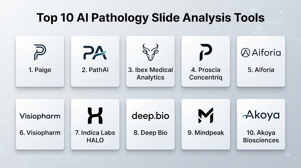
Top 10 AI Pathology Slide Analysis Tools
1 — Paige
One-line verdict: Best for pathology organizations seeking AI-assisted cancer detection and computational pathology within digital pathology workflows.
Short description:
Paige develops AI technologies for pathology, with a strong emphasis on cancer detection and computational pathology. Its solutions are designed to help pathologists analyze digital tissue images and identify clinically relevant patterns.
Standout Capabilities
- AI-assisted pathology analysis
- Cancer detection support
- Digital pathology workflows
- Tissue-image analysis
- Computational pathology
- Pathologist decision support
- Quantitative analysis
- Research-oriented pathology AI
AI-Specific Depth
- Model support: Primarily proprietary pathology AI models.
- RAG / knowledge integration: Primarily image-based computational pathology rather than conventional document RAG.
- Evaluation: Product-specific clinical validation varies according to the intended use.
- Guardrails: Human pathologist review and defined clinical indications are important.
- Observability: Product-specific operational monitoring varies.
Pros
- Strong specialization in computational pathology.
- Focused on clinically relevant pathology applications.
- Relevant to organizations developing digital pathology programs.
Cons
- Individual applications require separate clinical evaluation.
- Digital-slide infrastructure is generally required.
- Advanced pathology AI requires careful validation across laboratories.
Security & Compliance
Healthcare organizations should verify applicable regulatory authorization, encryption, access controls, audit logging, retention, residency, and certifications for the specific product and deployment.
Deployment & Platforms
- Web: Digital pathology workflow dependent
- Windows/macOS/Linux: Integration varies
- iOS/Android: Varies
- Deployment: Cloud and integrated options vary
Integrations & Ecosystem
- Whole-slide imaging
- Digital pathology viewers
- Pathology workflows
- Laboratory systems
- Scanner infrastructure
- Research environments
- Clinical applications
Pricing Model
Enterprise/custom pricing. Exact pricing varies by application and deployment.
Best-Fit Scenarios
- Cancer centers
- Digital pathology programs
- Pathology departments implementing AI-assisted diagnosis
2 — Ibex Medical Analytics
One-line verdict: Best for pathology laboratories seeking AI-assisted diagnosis and quality support across multiple tissue and disease workflows.
Short description:
Ibex Medical Analytics develops AI systems for digital pathology, focusing on assisting pathologists with detection and characterization of selected diseases and findings. Its platform approach is relevant to laboratories seeking broader pathology-AI capabilities.
Standout Capabilities
- Digital pathology analysis
- Cancer detection
- Tissue classification
- Pathologist assistance
- Multiple pathology applications
- Quality support
- Automated image analysis
- Clinical workflow integration
AI-Specific Depth
- Model support: Proprietary pathology AI models.
- RAG / knowledge integration: Primarily image-based analysis.
- Evaluation: Product-specific clinical evidence varies.
- Guardrails: Human pathologist review is central to appropriate use.
- Observability: Product-specific monitoring varies.
Pros
- Broad pathology-AI orientation.
- Designed around pathologist workflows.
- Supports multiple potential pathology applications.
Cons
- Individual algorithms require independent assessment.
- Performance can depend on slide quality and laboratory conditions.
- Deployment requires digital pathology infrastructure.
Security & Compliance
Verify regulatory authorization, encryption, access controls, data retention, residency, audit logs, and applicable certifications for each intended deployment.
Deployment & Platforms
- Web: Digital pathology workflow dependent
- Windows/macOS/Linux: Integration varies
- iOS/Android: Varies
- Deployment: Cloud/local options vary
Integrations & Ecosystem
- Whole-slide scanners
- Digital pathology viewers
- Laboratory systems
- LIS
- Pathology workstations
- Clinical workflows
- Research environments
Pricing Model
Enterprise/custom pricing. Exact pricing varies.
Best-Fit Scenarios
- Large pathology laboratories
- Cancer centers
- Multi-site pathology networks
3 — PathAI
One-line verdict: Best for organizations combining computational pathology, biomarker analysis, pharmaceutical research, and clinical development workflows.
Short description:
PathAI focuses on artificial intelligence for pathology and has applications spanning research, drug development, biomarker analysis, and computational pathology. Its capabilities are particularly relevant to pharmaceutical and research organizations as well as selected clinical environments.
Standout Capabilities
- Computational pathology
- Biomarker analysis
- Tissue image analysis
- Drug-development support
- Clinical research
- Quantitative pathology
- Image-based phenotyping
- Pathology research
AI-Specific Depth
- Model support: Proprietary computational pathology models and application-specific systems.
- RAG / knowledge integration: Primarily image analysis; broader data integration varies.
- Evaluation: Research and application-specific evaluation varies.
- Guardrails: Human expert review and study-specific validation are important.
- Observability: Monitoring capabilities vary by deployment.
Pros
- Strong research orientation.
- Relevant to pharmaceutical and clinical-trial workflows.
- Broad computational pathology capabilities.
Cons
- Some capabilities are more research-oriented than routine clinical diagnosis.
- Clinical deployment requirements vary by application.
- Advanced workflows can require specialized expertise.
Security & Compliance
Organizations should verify security controls, data processing, retention, residency, access management, auditability, and relevant certifications for the specific use case.
Deployment & Platforms
- Web: Varies
- Windows/macOS/Linux: Research and enterprise integration varies
- iOS/Android: Varies
- Deployment: Cloud and custom enterprise options vary
Integrations & Ecosystem
- Whole-slide imaging
- Digital pathology
- Research databases
- Pharmaceutical workflows
- Clinical-trial environments
- Biomarker workflows
- Computational pathology systems
Pricing Model
Enterprise/custom pricing.
Best-Fit Scenarios
- Pharmaceutical companies
- Contract research organizations
- Academic pathology research
4 — Aiforia
One-line verdict: Best for organizations seeking flexible AI-powered tissue image analysis across pathology research and selected clinical applications.
Short description:
Aiforia provides cloud-based AI-powered image analysis for digital pathology and related biological imaging applications. Its platform can support organizations that need to develop or apply image-analysis models across different tissue types.
Standout Capabilities
- Tissue image analysis
- Cell detection
- Cell classification
- Tissue segmentation
- Quantitative analysis
- Model development
- Digital pathology workflows
- Research applications
AI-Specific Depth
- Model support: AI image-analysis models with platform-level customization capabilities.
- RAG / knowledge integration: Not primarily a RAG platform.
- Evaluation: Model evaluation capabilities depend on the specific workflow.
- Guardrails: Human review and application-specific validation are required.
- Observability: Platform and workflow monitoring vary.
Pros
- Flexible image-analysis capabilities.
- Strong quantitative pathology orientation.
- Useful for research and specialized workflows.
Cons
- Requires users to understand image-analysis workflows.
- Clinical deployment requires appropriate validation.
- Model development can require specialized expertise.
Security & Compliance
Verify applicable security controls, encryption, access management, data retention, residency, auditability, and certifications.
Deployment & Platforms
- Web: Yes
- Windows/macOS/Linux: Browser and integration dependent
- iOS/Android: Varies
- Deployment: Cloud-oriented
Integrations & Ecosystem
- Whole-slide images
- Digital pathology
- Research systems
- Image-analysis workflows
- Laboratory environments
- Quantitative pathology
- Scientific imaging
Pricing Model
Subscription and enterprise/custom structures may vary by deployment and usage.
Best-Fit Scenarios
- Pathology research
- Pharmaceutical research
- Organizations needing customizable image analysis
5 — Deep Bio
One-line verdict: Best for organizations exploring AI-powered computational pathology and biomarker discovery in research-intensive environments.
Short description:
Deep Bio develops AI technologies focused on pathology and cancer-related image analysis. Its work is relevant to organizations exploring computational pathology, cancer detection, and quantitative tissue analysis.
Standout Capabilities
- Computational pathology
- Cancer image analysis
- Tissue classification
- Quantitative pathology
- AI-assisted pathology
- Research workflows
- Digital pathology
- Biomarker-oriented analysis
AI-Specific Depth
- Model support: Proprietary pathology AI.
- RAG / knowledge integration: Primarily image-based.
- Evaluation: Product-specific evidence varies.
- Guardrails: Human expert review remains important.
- Observability: Product-specific monitoring varies.
Pros
- Strong computational pathology orientation.
- Relevant to cancer-focused analysis.
- Suitable for research-intensive environments.
Cons
- Product scope varies by application.
- Clinical deployment requires indication-specific validation.
- Digital pathology infrastructure is required.
Security & Compliance
Verify applicable regulatory status, security architecture, encryption, data retention, residency, access controls, and certifications.
Deployment & Platforms
- Web: Varies
- Windows/macOS/Linux: Integration varies
- iOS/Android: Varies
- Deployment: Cloud/integrated options vary
Integrations & Ecosystem
- Whole-slide imaging
- Digital pathology
- Pathology systems
- Research workflows
- Cancer analysis
- Image-analysis platforms
Pricing Model
Enterprise/custom pricing. Exact pricing varies.
Best-Fit Scenarios
- Cancer research
- Digital pathology programs
- Computational pathology projects
6 — Proscia
One-line verdict: Best for pathology organizations seeking digital pathology infrastructure combined with computational pathology and AI capabilities.
Short description:
Proscia develops digital pathology software and computational pathology technologies designed to support pathology laboratories and healthcare organizations. Its ecosystem combines digital slide management with AI-oriented pathology capabilities.
Standout Capabilities
- Digital pathology
- Whole-slide image management
- Computational pathology
- AI-assisted analysis
- Pathology workflow support
- Image viewing
- Case management
- Enterprise pathology infrastructure
AI-Specific Depth
- Model support: Proprietary and ecosystem-based pathology AI capabilities vary.
- RAG / knowledge integration: Primarily digital pathology and image analysis.
- Evaluation: Product-specific validation varies.
- Guardrails: Pathologist review remains central.
- Observability: Enterprise monitoring capabilities vary.
Pros
- Strong digital pathology foundation.
- Relevant to enterprise pathology workflows.
- Combines infrastructure and AI-oriented capabilities.
Cons
- Enterprise deployment can be complex.
- Individual AI applications require separate evaluation.
- Organizations need to assess interoperability with existing pathology systems.
Security & Compliance
Verify encryption, access controls, audit logs, retention, residency, regulatory status, and applicable certifications for the specific deployment.
Deployment & Platforms
- Web: Yes
- Windows/macOS/Linux: Enterprise integration varies
- iOS/Android: Varies
- Deployment: Cloud and enterprise options vary
Integrations & Ecosystem
- Whole-slide scanners
- Digital pathology viewers
- LIS
- Pathology workflows
- AI applications
- Laboratory systems
- Enterprise imaging
Pricing Model
Enterprise/custom pricing.
Best-Fit Scenarios
- Enterprise pathology laboratories
- Digital pathology transformation
- Multi-site pathology organizations
7 — Paige AI
One-line verdict: Best for pathology teams prioritizing AI-assisted cancer detection and digital-slide interpretation support.
Short description:
Paige’s pathology AI technologies focus on computational analysis of tissue images, particularly cancer-related applications. Its systems are designed to provide additional information to pathologists working with digital slides.
Standout Capabilities
- Cancer detection
- Digital pathology
- Tissue analysis
- AI-assisted interpretation
- Computational pathology
- Quantitative analysis
- Pathologist workflow support
- Research applications
AI-Specific Depth
- Model support: Proprietary pathology AI.
- RAG / knowledge integration: Primarily image-based.
- Evaluation: Application-specific clinical validation varies.
- Guardrails: Human pathologist oversight is essential.
- Observability: Product-specific monitoring varies.
Pros
- Strong pathology specialization.
- Focus on clinically meaningful applications.
- Relevant to cancer-focused digital pathology workflows.
Cons
- Product-specific capabilities vary.
- Clinical deployment requires appropriate validation.
- Digital pathology infrastructure is necessary.
Security & Compliance
Verify applicable regulatory authorization, data controls, encryption, retention, residency, auditability, and certifications.
Deployment & Platforms
- Web: Digital pathology workflow dependent
- Windows/macOS/Linux: Integration varies
- iOS/Android: Varies
- Deployment: Cloud/integrated options vary
Integrations & Ecosystem
- Whole-slide imaging
- Pathology viewers
- Digital pathology systems
- Laboratory workflows
- Research environments
- Clinical pathology
Pricing Model
Enterprise/custom pricing.
Best-Fit Scenarios
- Cancer centers
- Digital pathology programs
- Pathology departments adopting AI-assisted analysis
8 — HistoWiz
One-line verdict: Best for research organizations seeking integrated digital pathology services and AI-supported tissue-image analysis.
Short description:
HistoWiz provides digital pathology and histology-related services, supporting researchers with tissue preparation, digitization, and image analysis workflows. Its relevance is strongest in research and life-sciences environments rather than as a universal clinical diagnostic platform.
Standout Capabilities
- Histology workflows
- Slide digitization
- Whole-slide imaging
- Image analysis
- Research pathology
- Quantitative tissue analysis
- Laboratory services
- Digital pathology workflows
AI-Specific Depth
- Model support: Application-specific AI and image-analysis capabilities vary.
- RAG / knowledge integration: Not primarily a RAG system.
- Evaluation: Research-specific evaluation varies.
- Guardrails: Human scientific review and study-specific validation are important.
- Observability: Workflow monitoring varies.
Pros
- Strong research orientation.
- Can combine physical histology and digital imaging workflows.
- Useful for organizations without extensive in-house pathology infrastructure.
Cons
- Not primarily a general-purpose clinical pathology AI platform.
- Capabilities depend on the research workflow.
- Clinical diagnostic use should not be assumed.
Security & Compliance
Research organizations should verify applicable data-handling, security, access, retention, and regulatory requirements for their specific projects.
Deployment & Platforms
- Web: Yes
- Windows/macOS/Linux: Workflow dependent
- iOS/Android: Varies
- Deployment: Service/cloud-oriented
Integrations & Ecosystem
- Histology
- Whole-slide imaging
- Research databases
- Image analysis
- Pharmaceutical research
- Scientific workflows
Pricing Model
Service-based and custom pricing. Exact pricing varies.
Best-Fit Scenarios
- Academic research
- Pharmaceutical research
- Contract research workflows
9 — Ibex Galen Platform
One-line verdict: Best for pathology laboratories seeking integrated AI assistance across multiple diagnostic pathology workflows.
Short description:
The Galen platform from Ibex is designed around AI-assisted pathology analysis. Its focus is helping pathologists identify and characterize selected findings across digital pathology workflows.
Standout Capabilities
- AI-assisted pathology
- Cancer detection
- Tissue analysis
- Cell-level analysis
- Diagnostic support
- Digital pathology integration
- Multiple pathology applications
- Workflow assistance
AI-Specific Depth
- Model support: Proprietary pathology AI models.
- RAG / knowledge integration: Primarily image analysis.
- Evaluation: Clinical validation varies by indication.
- Guardrails: Human pathologist review and workflow controls.
- Observability: Product-specific monitoring varies.
Pros
- Strong clinical pathology orientation.
- Broad application potential.
- Designed around pathologist workflows.
Cons
- Individual use cases require independent assessment.
- Requires suitable digital pathology infrastructure.
- AI outputs require professional interpretation.
Security & Compliance
Verify current regulatory authorization, encryption, access controls, audit logging, data retention, residency, and certifications.
Deployment & Platforms
- Web: Yes, depending on workflow
- Windows/macOS/Linux: Integration varies
- iOS/Android: Varies
- Deployment: Cloud/integrated options vary
Integrations & Ecosystem
- Whole-slide scanners
- Digital pathology
- LIS
- Pathology viewers
- Laboratory systems
- Clinical workflows
- AI applications
Pricing Model
Enterprise/custom pricing.
Best-Fit Scenarios
- Large pathology laboratories
- Cancer centers
- Multi-site pathology networks
10 — Visiopharm
One-line verdict: Best for organizations seeking advanced quantitative image analysis, tissue segmentation, and computational pathology workflows.
Short description:
Visiopharm develops image-analysis software for digital pathology and tissue research. Its technology is particularly relevant to organizations needing detailed quantitative analysis of cells, tissue structures, biomarkers, and spatial patterns.
Standout Capabilities
- Tissue segmentation
- Cell detection
- Cell classification
- Biomarker quantification
- Spatial analysis
- Quantitative pathology
- Image analysis
- Research workflows
AI-Specific Depth
- Model support: AI-based image-analysis capabilities with application-specific model workflows.
- RAG / knowledge integration: Primarily image-based computational analysis.
- Evaluation: Model-specific evaluation varies.
- Guardrails: Human review and study-specific validation are important.
- Observability: Platform monitoring varies.
Pros
- Strong quantitative analysis capabilities.
- Useful for complex tissue-analysis workflows.
- Relevant to research and pharmaceutical applications.
Cons
- Requires specialized pathology/image-analysis expertise.
- Clinical diagnostic use depends on the specific application.
- Advanced workflows can require substantial configuration.
Security & Compliance
Verify product-specific security, data handling, access control, retention, residency, and applicable regulatory requirements.
Deployment & Platforms
- Web: Varies
- Windows/macOS/Linux: Desktop and enterprise options vary
- iOS/Android: Varies
- Deployment: Local/cloud options vary
Integrations & Ecosystem
- Whole-slide images
- Digital pathology
- Image-analysis workflows
- Research databases
- Biomarker analysis
- Pharmaceutical research
- Laboratory systems
Pricing Model
Enterprise/custom pricing.
Best-Fit Scenarios
- Computational pathology research
- Pharmaceutical research
- Quantitative tissue analysis
Comparison Table
| Tool | Best For | Deployment | Model Flexibility | Strength | Watch-Out | Public Rating |
|---|---|---|---|---|---|---|
| Paige | Cancer-focused pathology AI | Cloud / integrated varies | Proprietary | Cancer detection support | Indication-specific validation | N/A |
| Ibex Medical Analytics | Clinical pathology AI | Cloud / integrated varies | Proprietary | Multi-application pathology support | Requires digital pathology infrastructure | N/A |
| PathAI | Research and biomarker analysis | Cloud / enterprise varies | Proprietary | Computational pathology | Strong research orientation | N/A |
| Aiforia | Flexible tissue analysis | Cloud-oriented | Customizable AI workflows | Quantitative image analysis | Requires expertise | N/A |
| Deep Bio | Cancer computational pathology | Integrated / varies | Proprietary | Cancer-focused analysis | Product scope varies | N/A |
| Proscia | Enterprise digital pathology | Cloud / enterprise | Ecosystem-dependent | Digital pathology infrastructure | Implementation complexity | N/A |
| Paige AI | Digital-slide cancer analysis | Cloud / integrated | Proprietary | Pathologist assistance | Application-specific scope | N/A |
| HistoWiz | Research pathology | Service / cloud-oriented | Application-dependent | Histology plus digital analysis | Primarily research-oriented | N/A |
| Ibex Galen | Diagnostic pathology workflows | Cloud / integrated | Proprietary | Pathology workflow support | Clinical validation by indication | N/A |
| Visiopharm | Quantitative computational pathology | Local / cloud varies | AI/custom workflows | Tissue and cell analysis | Specialized expertise required | N/A |
Scoring & Evaluation
The scores below are comparative editorial assessments rather than clinical accuracy ratings. They should not be interpreted as evidence that one system is clinically safer or diagnostically superior.
Actual procurement decisions should prioritize the exact intended use, regulatory authorization, clinical evidence, local validation, scanner compatibility, workflow integration, and patient-safety requirements.
| Tool | Core | Reliability/Eval | Guardrails | Integrations | Ease | Perf/Cost | Security/Admin | Support | Weighted Total |
|---|---|---|---|---|---|---|---|---|---|
| Paige | 9.2 | 9.0 | 9.0 | 8.9 | 8.3 | 8.0 | 9.0 | 8.7 | 8.8 |
| Ibex Medical Analytics | 9.2 | 9.0 | 9.0 | 9.0 | 8.3 | 8.0 | 9.0 | 8.7 | 8.8 |
| PathAI | 9.1 | 9.2 | 8.9 | 8.8 | 8.0 | 8.0 | 8.9 | 9.0 | 8.7 |
| Aiforia | 9.0 | 8.8 | 8.5 | 8.6 | 8.2 | 8.3 | 8.6 | 8.5 | 8.6 |
| Deep Bio | 8.7 | 8.8 | 8.6 | 8.4 | 8.1 | 8.0 | 8.5 | 8.3 | 8.4 |
| Proscia | 9.1 | 8.8 | 8.9 | 9.2 | 8.1 | 7.8 | 9.1 | 8.8 | 8.7 |
| Paige AI | 9.1 | 9.0 | 9.0 | 8.8 | 8.3 | 8.0 | 9.0 | 8.7 | 8.8 |
| HistoWiz | 8.2 | 8.2 | 8.0 | 7.9 | 8.3 | 7.8 | 8.0 | 8.3 | 8.1 |
| Ibex Galen | 9.2 | 9.0 | 9.0 | 9.0 | 8.3 | 8.0 | 9.0 | 8.7 | 8.8 |
| Visiopharm | 8.9 | 8.7 | 8.5 | 8.5 | 7.9 | 8.2 | 8.5 | 8.6 | 8.5 |
Top 3 for Enterprise
- Ibex Medical Analytics — strong fit for pathology laboratories seeking multiple AI-assisted diagnostic workflows.
- Paige — particularly relevant to cancer-focused digital pathology programs.
- Proscia — suitable for organizations prioritizing enterprise digital pathology infrastructure alongside computational pathology.
Top 3 for SMB
- Aiforia — attractive for focused image-analysis and quantitative pathology projects.
- Deep Bio — relevant for organizations with cancer-focused computational pathology needs.
- Visiopharm — useful for targeted quantitative tissue-analysis workflows.
Top 3 for Developers
- Aiforia — useful where customized image-analysis workflows are important.
- Visiopharm — relevant for sophisticated computational pathology analysis.
- PathAI — particularly relevant to research and computational pathology environments.
Which AI Pathology Slide Analysis Tool Is Right for You?
Solo / Freelancer
Individual pathologists generally should not independently deploy AI pathology systems for diagnostic purposes without institutional governance and appropriate clinical validation.
The key questions should be:
- Is the system intended for the specific clinical task?
- Is it appropriately authorized?
- Does it work with the laboratory’s scanner?
- Does it integrate with the pathology viewer?
- Can the pathologist review the evidence?
- Can the pathologist override the AI?
- How is patient information protected?
For research or educational use, the evaluation criteria can be different, with greater emphasis on flexibility and experimentation.
SMB
Smaller pathology laboratories should avoid starting with a complicated AI ecosystem.
Choose one high-value application.
Examples include:
- Cancer detection
- Immunohistochemistry quantification
- Tumor measurement
- Cell counting
- Quality control
Before purchasing, determine whether the laboratory already has:
- Whole-slide scanners
- Digital pathology viewers
- Storage infrastructure
- LIS integration
- Suitable network capacity
- Pathologist digital workflows
If these components are missing, digital pathology infrastructure may need to be addressed before AI provides meaningful value.
Mid-Market
Mid-sized laboratories can benefit from creating a formal computational pathology strategy.
Establish:
- AI governance
- Clinical ownership
- Technical ownership
- Validation standards
- Scanner compatibility standards
- Data-management policies
- Model monitoring
- AI inventory
- Version control
- Security review
- Incident handling
Avoid allowing individual departments to adopt unrelated AI systems without centralized oversight.
Enterprise
Large pathology networks should think beyond individual algorithms.
Enterprise requirements can include:
- Multi-site slide management
- Multiple scanner vendors
- Centralized AI governance
- Model inventory
- Workflow orchestration
- LIS integration
- Digital pathology interoperability
- Centralized security
- Performance monitoring
- Data residency
- Access management
- Auditability
- Disaster recovery
- Vendor management
- Long-term data retention
Enterprise pathology organizations should also consider whether a platform can support future AI applications without requiring a complete infrastructure redesign.
Regulated Industries
Pathology is particularly sensitive because AI can potentially influence cancer diagnosis, treatment decisions, clinical trials, and patient management.
Organizations should verify:
- Intended use
- Regulatory authorization
- Clinical validation
- Patient population
- Scanner compatibility
- Staining protocols
- Data processing
- Data retention
- Data residency
- Encryption
- Access controls
- Audit trails
- Vendor access
- Model updates
- Incident response
- Human oversight
Particular attention should be paid to laboratory-specific variables.
A model that performs well on slides generated by one scanner or staining workflow may behave differently under another environment.
Budget vs Premium
The cost of pathology AI extends beyond software licensing.
Organizations should calculate:
- Scanner costs
- Slide digitization
- Storage
- Network bandwidth
- AI processing
- Cloud infrastructure
- Software licensing
- Integration
- Validation
- Training
- Support
- Monitoring
- Security
- Laboratory workflow changes
Whole-slide images can be large, making storage and data-transfer requirements especially important.
A low-cost AI application may become expensive if it requires significant infrastructure changes.
Build vs Buy
Building pathology AI internally can be attractive for academic medical centers and research organizations.
A custom system may require:
- Whole-slide image processing
- Tissue segmentation
- Annotation tools
- Model training
- GPU infrastructure
- Data pipelines
- Validation
- Scanner normalization
- Stain normalization
- Model monitoring
- Clinical governance
- Security
- Regulatory strategy
For routine clinical deployment, buying an appropriately validated solution is often more practical.
Research institutions may reasonably choose a hybrid approach:
Commercial digital pathology infrastructure + internally developed research models.
Implementation Playbook
First 30 Days: Pilot + Success Metrics
Choose a single pathology workflow.
Examples:
- Prostate cancer detection
- Breast cancer analysis
- Immunohistochemistry quantification
- Tumor segmentation
- Cell counting
- Quality control
Establish a baseline.
Measure:
- Number of slides
- Average review time
- Manual counting time
- Diagnostic turnaround time
- Existing disagreement rate
- Number of cases requiring additional review
- Slide-quality problems
- Digitization time
Create a representative evaluation dataset.
Include:
- Normal cases
- Positive cases
- Borderline cases
- Difficult cases
- Poor-quality slides
- Different staining conditions
- Different scanners
- Different laboratories where applicable
Days 31–60: Security + Evaluation + Rollout
Perform technical and clinical validation.
Evaluate:
- Sensitivity
- Specificity
- False-positive rate
- False-negative rate
- Processing latency
- Slide-failure rate
- Pathologist acceptance
- Workflow impact
- Scanner compatibility
Test different image conditions.
Pay attention to:
- Staining variability
- Tissue artifacts
- Folded tissue
- Out-of-focus regions
- Tissue fragmentation
- Compression
- Scanner differences
- Low-quality scans
Security evaluation should include:
- Authentication
- Authorization
- Encryption
- Access logging
- Data retention
- Data residency
- Vendor access
- Network architecture
- API security
- Incident response
Days 61–90: Cost, Latency, Governance + Scale
After successful validation:
- Expand to additional pathologists.
- Increase case volume gradually.
- Monitor AI performance.
- Track model versions.
- Measure processing costs.
- Monitor storage requirements.
- Review false positives.
- Review false negatives.
- Measure pathologist workload.
- Establish AI governance.
- Define update procedures.
- Establish incident-management processes.
- Create retirement criteria.
At scale, monitor whether AI performance changes across:
- Scanner models
- Laboratories
- Staining protocols
- Patient populations
- Tissue types
- Disease prevalence
- Model versions
Common Mistakes & How to Avoid Them
- Treating AI output as a diagnosis: Keep the pathologist responsible for final clinical interpretation.
- Ignoring slide quality: Poor digitization can negatively affect AI performance.
- Ignoring scanner variation: Different scanners can produce different image characteristics.
- Ignoring staining variation: Histology and immunohistochemistry staining differences can affect model performance.
- Skipping local validation: External validation does not guarantee identical local performance.
- Using research models clinically without appropriate validation: Research capability does not automatically establish clinical suitability.
- Ignoring false positives: Excessive AI findings can increase pathologist workload.
- Ignoring false negatives: Missed pathology findings can have significant clinical implications.
- No human review: AI should operate within an appropriate pathologist workflow.
- Poor digital pathology integration: AI should fit naturally into the existing slide-review environment.
- Ignoring storage costs: Whole-slide images can require substantial storage.
- Ignoring network bandwidth: Large digital slides can create significant data-transfer requirements.
- No model-version tracking: Updates can alter performance.
- No scanner compatibility testing: AI should be evaluated with the actual scanning infrastructure.
- Ignoring bias: Performance may differ across populations, laboratories, scanners, and staining methods.
- No monitoring after deployment: Clinical performance can change over time.
- No fallback workflow: Pathology operations must continue when AI or digital infrastructure is unavailable.
- Over-automating diagnostic decisions: AI should not eliminate appropriate professional review.
- Ignoring interoperability: Digital pathology systems need reliable communication between scanners, viewers, LIS, and AI.
- Choosing based on demonstrations: Demonstration slides rarely represent the full complexity of routine pathology.
- Ignoring governance: Every clinical AI model should have clear ownership, validation, monitoring, and retirement processes.
FAQs
What is AI Pathology Slide Analysis?
AI Pathology Slide Analysis uses artificial intelligence to examine digitized pathology slides and assist with tasks such as tissue classification, cell detection, cancer identification, biomarker quantification, and quantitative analysis.
What is a whole-slide image?
A whole-slide image is a high-resolution digital representation of an entire pathology slide. It allows the tissue to be viewed digitally instead of relying exclusively on a glass slide and microscope.
Can AI diagnose cancer from pathology slides?
Some AI systems are designed and authorized for specific pathology-related clinical applications. However, the appropriate role of each system depends on its intended use, validation, regulatory status, and clinical workflow.
Can AI replace pathologists?
AI should not be treated as a general replacement for pathologists. Pathologists provide clinical interpretation, contextual reasoning, quality assessment, and final diagnostic responsibility.
What types of pathology can AI analyze?
Depending on the system, AI can support histopathology, immunohistochemistry, oncology, cytology-related workflows, tissue classification, and other specialized applications.
Can AI analyze immunohistochemistry slides?
Yes, some AI systems support quantitative analysis of selected immunohistochemistry biomarkers. The exact markers and intended uses vary by product.
What is computational pathology?
Computational pathology uses digital pathology images, image analysis, machine learning, and other computational techniques to extract quantitative and qualitative information from tissue.
What is the difference between digital pathology and AI pathology?
Digital pathology refers to the digitization and digital management of pathology slides and workflows. AI pathology adds algorithms that analyze those digital images.
Does AI pathology require a whole-slide scanner?
For many whole-slide AI workflows, digital slides are required. However, specific applications may have different image-input requirements.
Can AI work with different pathology scanners?
Compatibility varies. Organizations should test the specific AI application with the scanner models and image formats used in their laboratory.
Why is local validation important?
Pathology images can differ because of scanners, staining protocols, tissue preparation, patient populations, laboratories, and image quality. Local validation helps determine whether an AI system performs appropriately in the intended environment.
Can AI analyze multiple tissue types?
Some platforms support multiple tissue types, while others specialize in particular organs or diseases. Capabilities vary by product.
What is tissue segmentation?
Tissue segmentation involves identifying and separating different regions or structures within a pathology image, such as tumor, stroma, necrosis, or other tissue components.
What is cell segmentation?
Cell segmentation identifies individual cells or cell boundaries in an image so that the system can count or characterize them.
Can AI count cells?
Yes. Selected pathology AI systems can identify and count cells, although accuracy depends on the tissue type, staining, image quality, and specific application.
Can AI quantify biomarkers?
Some systems can quantify selected biomarkers, including certain immunohistochemistry measurements. The specific biomarker support varies by application.
What security issues should pathology laboratories consider?
Important areas include encryption, authentication, authorization, audit logging, data retention, data residency, network security, vendor access, API security, and incident response.
Can pathology AI run in the cloud?
Yes, depending on the product. Cloud deployment can simplify infrastructure management but introduces additional considerations around privacy, security, network connectivity, latency, and data residency.
Can pathology AI run on-premises?
Some products and architectures support local deployment, while others are cloud-oriented. Deployment options should be verified for the specific application.
How much storage does AI pathology require?
Whole-slide images can be large, so storage requirements depend on slide resolution, compression, number of slides, retention period, and the organization’s workflow.
Does AI pathology require GPUs?
Some AI workflows require substantial computational resources, while others are optimized for cloud or managed infrastructure. Requirements vary by application and deployment model.
How should a pathology AI model be evaluated?
Evaluate sensitivity, specificity, false positives, false negatives, robustness, scanner compatibility, staining variability, patient populations, workflow impact, processing latency, and clinical usefulness.
What is model drift in pathology AI?
Model drift occurs when changes in clinical or technical conditions cause an AI system’s real-world performance to differ from its original validation performance.
Why does staining matter for AI?
Differences in staining intensity, preparation, reagents, and laboratory protocols can alter image characteristics and potentially affect model performance.
What happens if the AI system fails?
The laboratory should maintain a fallback workflow that allows pathologists to continue reviewing cases without relying entirely on AI availability.
Can pathology AI be used for research?
Yes. Research is an important area for computational pathology, including biomarker discovery, drug development, tissue analysis, and clinical research.
Can pharmaceutical companies use pathology AI?
Yes. Computational pathology can support drug-development research, biomarker analysis, clinical trials, and quantitative tissue assessment.
Is open-source pathology AI suitable for clinical diagnosis?
Open-source models can be valuable for research, but clinical deployment requires appropriate validation, governance, security, quality controls, and regulatory consideration.
Can hospitals build their own pathology AI?
Yes, particularly research hospitals with access to high-quality pathology datasets and specialized AI teams. Clinical deployment requires considerably more than model development.
What is the biggest mistake when purchasing pathology AI?
Choosing a system based solely on reported accuracy without evaluating local scanner compatibility, staining variation, patient population, clinical validation, workflow integration, and ongoing monitoring.
How should pathologists interact with AI?
AI should provide useful evidence and analysis within the pathologist’s normal workflow, allowing the professional to review findings, interpret context, and make the final clinical decision.
Conclusion
AI Pathology Slide Analysis is becoming an important component of computational pathology and digital pathology. Its potential extends well beyond simple image classification.Paige and Ibex Medical Analytics are particularly relevant to clinical pathology AI and cancer-focused applications. PathAI is highly relevant to computational pathology, pharmaceutical research, and biomarker analysis. Aiforia and Visiopharm are strong considerations for flexible quantitative image-analysis workflows. Proscia is relevant to organizations building broader digital pathology infrastructure, while Deep Bio focuses on computational pathology and cancer analysis. HistoWiz is more research-oriented and can be useful where histology and digital analysis need to work together.